

Wisdom tooth surgery
August 28, 2020
Gum Disease
September 5, 2020
Tooth Infection Related To Dens Evagination
Prepared by Dr. Lim Ming Chau, Union Dental Bayan Baru.The appearance of teeth varies in sizes in different individuals but they have a typical shape to serve their specific functions. Premolar teeth have two cusps on the occlusal surface. These are the normal anatomy of premolars.
However, sometimes acquired and inherited developmental abnormalities may alter the size, shape and number of teeth. Dens evaginatus is a developmental anomaly characterised by projection or tubercle protuberance of enamel and dentine that usually encloses the pulp tissue. It occurs more frequently on premolars, which is also known as Leong’s premolar.
However, sometimes acquired and inherited developmental abnormalities may alter the size, shape and number of teeth. Dens evaginatus is a developmental anomaly characterised by projection or tubercle protuberance of enamel and dentine that usually encloses the pulp tissue. It occurs more frequently on premolars, which is also known as Leong’s premolar.
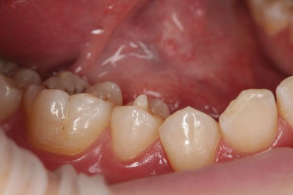
A projected tooth structure in between two cusps of premolar, known as Dens Evagination.

The accessory cusp is often prone to worn or fracture in normal function or trauma.

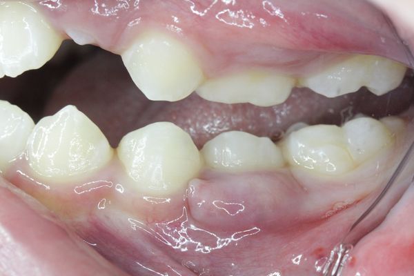
Image shown a premolar with worn Dens Evagination.
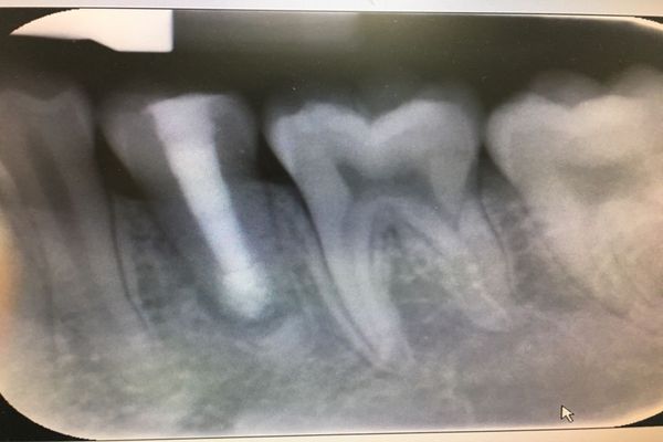
Dental Abscess & Pus Discharging Sinus
If no proper treatment is done on the fractured cusp, there will be chances for bacteria to infect the pulp of the tooth. It may lead to pulpal and periapical pathology such as abscess or sinus formation.
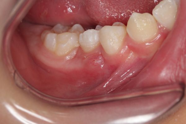
A pus discharging sinus tract related to infected premolar.
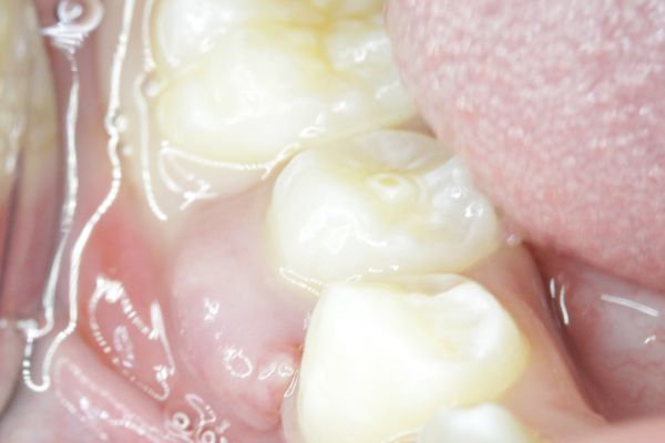
Dental abscess associated to the infected Premolar.
Treatment Protocols
For Healthy Pulp

Preventive Filling
Preventive treatment is needed to protect the tooth. In dens evaginatus of vital teeth, selective grinding of projected cusp and restoring with composite is the treatment of choice.
Preventive treatment is needed to protect the tooth. In dens evaginatus of vital teeth, selective grinding of projected cusp and restoring with composite is the treatment of choice.
For Necrotic Pulp

Root Canal Treatment
Root canal treatment is require to remove necrotic pulp tissue, disinfect the canal and sealed it to prevent bacteria invasion again.
Root canal treatment is require to remove necrotic pulp tissue, disinfect the canal and sealed it to prevent bacteria invasion again.
For Necrotic Pulp with Open Apex
Apexification
In non-vital teeth with incomplete root development, apexification with calcium hydroxide non-setting, Mineral Trioxide Aggregate ( MTA ) or Bio-ceramic Root Canal Sealer, to promote closure of the apex foramen by formation of cementum.
In non-vital teeth with incomplete root development, apexification with calcium hydroxide non-setting, Mineral Trioxide Aggregate ( MTA ) or Bio-ceramic Root Canal Sealer, to promote closure of the apex foramen by formation of cementum.
